

The MEIBOMIAN GLANDS
Links to pages:
GLIMPSE on …
The MEIBOMIAN GLANDS
SUMMARY (Every sentence here is LINKED to a respective CHAPTER below)
Tear Film LIPIDS are of utmost Importance for the Function of the TEAR FILM and thus for the Ocular Surface as a whole. The MEIBOMIAN GLANDS inside the eye lids produce lipids. The Meibomian Lipids form an OIL at body temperature. The Meibomian Oil forms the superficial phase of the tear FILM - The tear FILM LIPID LAYER (TFLL). The Meibomian OIL in the Tear Film has important functions for the STABILITY of the Tear Film. Meibomian oil RETARDS the EVAPORATION of the aqueous phase. Meibomian OIL provides a HOMOGENEOUS and perfectly SMOOTH SURFACE for the Tear FILM for perfect light refraction and visual acuity. The lipid production for the tear film is achieved by a large number of individual Meibomian glands. Every single Meibomian Gland has its own orifice. Meibomian Glands have some structural characteristics in contrast to other sebaceous glands such as a long slender gland body. Therefore, SECRETION and DELIVERY of the oil occur in separate regions in Meibomian Glands. SECRETION is the production of the oil deep down inside the Glands. DELIVERY on the other ´end´ describes the process of driving out or delivering the oil from the orifice onto the lid margin. The Differentiation between SECRETION and Delivery explains Obstruction as a main pathology of the Meibomian Glands.
Tear Film LIPIDS are of utmost Importance for the Function of the Tear Film and thus for the Ocular Surface as a whole.

The Oil from the Meibomian Glands inside the Eye Lids covers the aqueous Tear FILM with an oily layer that retards evaporation of water and increases the stability of the tear FILM
The Meibomian glands are lipid producing sebaceous glands in the eye lids. The lipids are very important for the homeostasis of the tear film and for vision.
The Tear Film Lipid Layer is the outer surface of the tear film, it improves tear film stability through retarding the evaporation of the aqueous main phase and provides a smooth homogeneous surface for refraction of incoming light.
Due to the important functions of the Meibomian Lipids for the TEAR FILM ... and since the tear film is the most important requirement for health and integrity of the Ocular Surface ... it is no surprise, that Meibomian Gland Dysfunction (MGD) is the main cause for Dry Eye Disease.
For more Details please see the Section on Ocular Surface and the Section on Tears
The Meibomian Glands inside the eye lids produce lipids in the form of a liquid clear oil
The meibomian lipids form an oil at body temperature

The Meibomian glands are located inside the tarsal plates of both the upper and lower eye lids.
They produce lipids that are liquid at body temperature and thus form an OIL.
... this note may already point to a potential issue in pathology
... because when the temperature inside the eye lids goes down or the composition of lipids may change (which can both occur) - the Meibomian lipids may change from a nice oil into a highly viscous solution or may even become solid - like olive oil will change its physical state from oily to solidification when it is accidentally stored outside in winter
Whether a gland actively produces normal liquid clear oil can be checked by a “diagnostic expression”
"DIAGNOSTIC Expression” of the Meibomian glands
Healthy Meibomian Glands deliver a clear liquid oil (arrow) through their orifice onto the lid margin. This can be tested by a technique termed ´Diagnostic Expression´ with very mild pressure from the outside - every orifice should then deliver a little puddle of oil (arrow).
If no small puddle of oil is visible at the gland openings, a simple test of the glandular activity can be performed as a precaution.
With the finger, a gentle pressure can be applied to the edge of the eyelid from the outside (see illustration).
This pressure should be of the same order of magnitude as the normal eyelid exerts by orbicularis muscle during eyelid closure.
This is called a "Diagnostic Expression" of the meibomian glands and goes back to Donald KORB, Boston.
A normally functioning Meibomian gland should then give off a small droplet of thin, clear secretion.
Meibomian oil forms the superficial phase of the Tear Film – the tear film lipid layer (TFLL)
The Tear Film in front of the transparent cornea (the pre-corneal tear film) is a layered structure in which typically three layers are discriminated: (1) at the outside surface to the ambient air is, as just explained in the text to the left, the thin layer of Meibomian Oil (yellow). The oil covers (2) the aqueous main phase of the tear film that is conceivably to some extent mixed with Mucins that constitute in a concentrated for the (3) inner Mucin (pink) Layer that is locate directly on and produced by the cells of the surface epithelium of the ocular surface. The thin lipid layer on top is in the schematic drawing shown much too broad - because, in the real size relation, it would be almost impossible to see it in cross section through a tear film.
The Meibomian oil covers the water-mucin main phase of the tear film as a surface layer - like a lid on a pot of warm water:
Meibomian oil constitutes the most superficial smooth tear film layer
that is termed the Tear Film Lipid Layer (often abbreviated as TFLL)
which represents the air-tear-interface
The meibomian oil in the tear film lipid layer (TFLL) has important functions for the stability of the Tear Film
Thereby the Meibomian lipids in the Tear Film Lipid Layer, are thought to have, among probably many others, at least two important functions as the surface layer of the pre-corneal tear film:

The main functions, among probably many others, are the retardation of evaporation of the aqueous phase, the water of the tear film, by sealing the tear film like a lid on a pot of warm water. A second important function is to provide a very smooth and homogeneous surface for the air-to-tear interface that is the main refractory surface of the eye. It provides about 3/4 of the refractive power of the eye.
(1) Meibomian oil RETARDS the EVAPORATION of the aqueous phase
at the open palpebral fissure of the ocular surface
the (warm) tear fluid is exposed to the environmental ambient air
this leads to a rapid evaporation of water
Pathological DEFICIENCY of the Meibomiman Oil results in Increased Evaporation, often termed HYPER-EVAPORATION, of the Tear Water. This condition is known as the lipid-deficient evaporative type of Dry Eye Disease - which is the main Type of Dry Eye
(2) Meibomian OIL provides a homogeneous and perfectly smooth surface for the Tear Film for perfect light refraction and visual acuity

it is the main refractory surface of the eye
responsible for about three quarters of the refractive power of the eye and
thus for visual acuity
It is therefore it is not surprising that Pathological Alterations of the homogeneity of the Tear Film Lipid Layer result in a loss of visual acuity - this is an early and often reported finding in tear deficiency during Dry Eye Disease. Since the visual acuity is often fluctuating with environmental conditions and the blink movement it is often termed ´unstable visual acuity´
Anatomy of the Meibomian Glands
The Meibomian Glands have a very characteristic Anatomy that differentiates them clearly from other sebaceous glands in the body ... and also offers some hints for potential issues that may be relevant for pathology
... why are they called glands in the plural form ?
The lipid production for the Tear Film is achieved by a large number of individual Meibomian glands:
they each have their own individual gland body
there are about 30 glands in each upper eye lid
and about 25 glands in each lower eye lid
altogether there are typically more than 50 individual Meibomian glands for each eye
every single gland has its own individual ORIFICE
that opens at the posterior lid margin
directly before the start of the Line of MARX (for more details, please see there)
still within, and thus encircled by, the cornified epidermis - which, as a matter of fact even enters the end of the excretory duct for a very short distance
The extended long gland bodies of the MEIBOMIAN GLANDS can be seen through the conjunctiva when the eye lid (here the lower one) is slightly everted. The individual orifices (indicated here by open arrows) are typically arranged in a single row, very close to the posterior lid border - immediately in front of the anterior rim of the tear meniscus.
so, again, with an eye on potential pathology, ... let´s hope that the normal process of keratinization and eventual cornification that produces anuclear cornified squames ... remains nice and quiet - because exaggerated keratinization could obstruct the narrow lumen of the gland orifice
therefore it is not surprising that indeed hyper-keratinization, together with increased viscosity of the oily secretum are indeed a main pathogenetic factors for Meibomian Gland DYSFUNCTION (MGD) - the main cause of Dry Eye Disease
Some anatomical characteristics of meibomian glands
Other sebaceous glands are typically associated with hairs, as e.g. all the hairs in the skin have associated sebaceous glands. Sebaceous glands, particularly those in the skin, are typically short and stout with a short excretory duct that delivers the produced oil into the hair follicle around the hair shaft and onto the skin.
Meibomian glands in contrast:
are not associated with hairs
have a long slender gland body
have an elaborated fine ductal system
with a long central duct and narrow connecting ductules
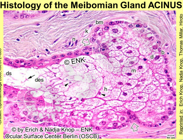
that lead to spherical secretory acini
acini are filled by secretory cells termed Meibocytes.
here again, some immediate thoughts come up as a link to potential pathology of the glands - a long, fine and slender ductal system that leads from deep in the tarsal plates to the surface of the posterior lid border ... is probably not ideal to drain an oily secretum that may easily change its viscosity depending on its composition and on the environmental weather conditions ...
... and these concerns unluckily meet closely what leads to and happens during Meibomian Gland Dysfunction - often better known by the abbreviation of MGD
Therefore, it does not come as a surprise, that
obstruction is the main pathology of the Meibomian glands and mainly their terminal ductal system, and the main therapeutic challenge is the removal of obstruction in order to get the lipid secretion flowing again.
What is a sebaceous gland ?
Sebaceous glands
produce an oily secretum
by formation and accumulation of lipids inside the secretory cells
this process is termed ´MATURATION´
during their maturation the cells move over several days
from the basal cell layer in the periphery of the acinus to the center
eventually the cells rupture and dissolve
basically all remnants of the whole cells form the Meibomian oil
Therefore sebaceous gland acini are completely filled by secretory cells. Since the secretory cells are completely lost during the secretory process they must be replenished by the similarly continuous cell division of stem cells
Secretion and Delivery of the Meibomian Glands – what is that ?
Due to the explained long extension of the Meibomian glands the secretory Meibocytes in the spherical acini are typically far away from the orifice at the posterior lid margin.
Therefore it makes sense to divide the process to provide oil onto the posterior lid margin and the tear film according to Bron and Tiffany into two steps that are separated in space and time:
SECRETION
describes the process of holocrine lipid production inside the acinus
it ends with the disruption of the secretory cells that forms the oily end product
that enters the start of the ductal system at the end of opening of the acinus
Secretion is a CONTINUOUS process because
it is driven by the continuous cell division and formation of new cells in the acinus
it thus results in a certain ´secretory pressure´ that conceivably also drives the oil within the ductal system towards the orifice
... then the secretum must be transported within the ductal system and eventually ...
DELIVERY
describes the process of driving out - or delivering - the oil
from the ductal system, through the orifice onto the lid margin
Delivery is a DIS-continuous process because
it needs the assistance of the lid muscles
is therefore linked to the blink movement of the eye lids
The differentiation between secretion and delivery allows the understanding of Gland Obstruction as a main pathology of the Meibomian Glands

In an obstructed gland (right) the delivery of the secreted oil is blocked and therefore the increasing pressure inside the gland leads to a dilatation of the ducts and to a pressure atrophy of the secretory acini.
The Differentiation between secretion deep down in the Gland that fills the ductal system with oil and the eventual delivery from the ductal system out of the gland onto the lid margin allows to better understand obstructive MGD.
Obstruction of the terminal part of a Meibomian Gland will necessarily lead to a stasis of Meibomian oil inside the gland and, since the secretion in the acini continues, the pressure inside the gland will increase. This will necessarily lead to the potential onset of pressure-dependent atrophy of the gland with dilatation of the ducts and destruction of the secretory tissue.